Double Floor Of Sella Turcica
Double Sellar Floor Sign A Clue Of Pituitary Tumor Springerlink
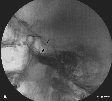
Double Sellar Floor Radiographic Sign For A Pituitary Adenoma Barrow

Specialised Projections Of The Skull Radiology Key

Magnification Of Lateral Cephalogram Case 2 The Sella Turcica Showed Download Scientific Diagram

Morphologic Classification Of Sella Turcica A Normal Sella Turcica Download Scientific Diagram

Sella Turcica X Ray Lateral View Legend No Evidence Of Mass Lesion Download Scientific Diagram
1 autopsy studies confirm the high disease prevalence reported to be 5 5 to 20 of the general population.
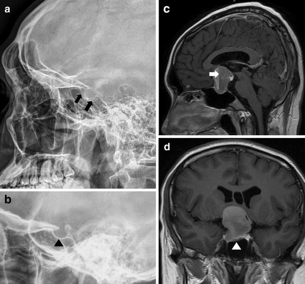
Double floor of sella turcica.

A Skull X Ray Showed A Double Floor Of Sella B C Magnetic Download Scientific Diagram

Morphological Variants Of Sella Turcica Seen In Lateral Cephalogram A Download Scientific Diagram

Enlarged Sella Turcica Differential Radiology Reference Article Radiopaedia Org

Full Text Association Of Sella Turcica Bridging With Palatal Canine Impaction In Ccide
Source : pinterest.com
